History:
A 75-year-old male suffered from impingement syndrome over left shoulder
Questions:
1. What are the findings?
2. What is the diagnosis?
Answers:
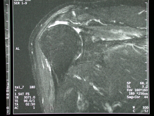
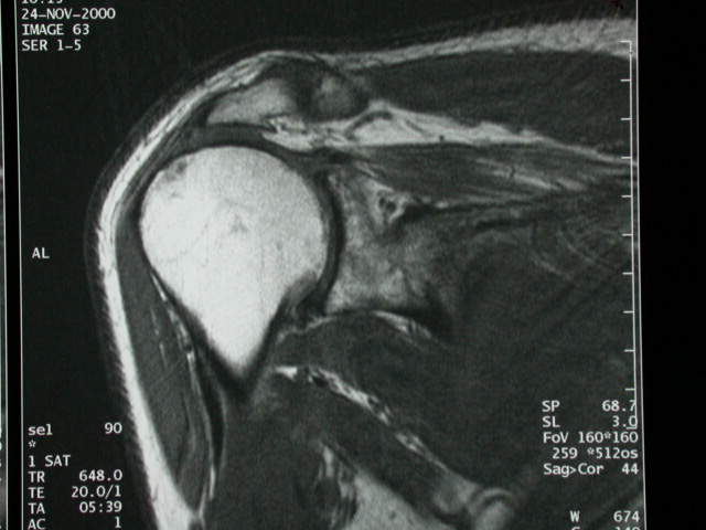
1. MRI of the left shoulder without contrast:
A complete tear in the supraspinatous tendon with joint fluid in
the gap between the torn edge of the supraspinatous tendon, extending
into the subdeltoid-subacromial space.
The acromioclavicular joint is degenerated and hypertrophic.
2. Acute full-thickness rotator cuff tear involving supraspinatous
tendon
Discussion
A full-thickness tear of the rotator cuff cause a gap in the tendon,
with the signal pattern of the gap related to the age of the tear.
At acute tear, in addition to edema, the gap between the torn edge
of the tendon fills with fluid as low signal intensity on T1-weighted
images and high signal intensity on T2-weighted images. Extension
into the subacromial-subdeltoid bursa can cause a massive bursal
effusion as a high signal structure on T2-weighted images. Extensive
tear can even continue into the acromioclavicular capsule to cause
an effusion of the acromioclavicular joint (the so-called "geyser'
sign"). Large effusion of the glenohumeral joint may be noted.
The defect in a chronic rotator cuff tear can fill with granulation
tissue (covered rupture) and remains isointense to hypointense on
T2-weighted images without obvious joint or bursal effusion. In
may be difficult to make the diagnosis at MRI and a confident distinction
from chronic tendinitis may be impossible.
Tears of the supraspinatus muscle occur most frequently as part
of the impingement syndrome, whereas tears of the infraspinatus
and subscapularis are less frequent and usually are traumatic in
origin.
MRI has proved a highly sensitive method for the detection of full-thickness
tear. The sensitivity has been found to be 80-100% and the specificity
higher than 90%, comparable to the results of conventional arthrography.
The sensitivity and specificity are considered less for partial
tears, chronic covered small tears and chronic tendinitis.
Reference
1. Kursunoglu-Brahme, S., D. Resnick: Magnetic resonance imaging
of the shoulder. Radiol. Clin. N. Amer. 28 (1990): 941-54 |


