History:
A 57 y/o male found a left popliteal mass noted for 6 months. There
is a history of previous trauma over left knee with pain.
Questions:
1. What are the findings?
2. What is the differential diagnosis?
3. What is the diagnosis?
Answers:
1. MRI of the left knee with/without contrast:
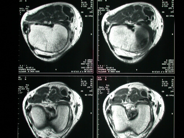
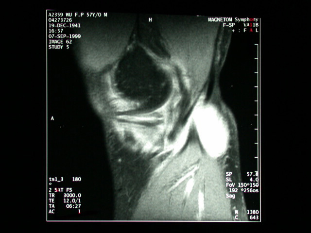
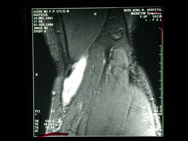
l A huge cystic lesion with thickened wall is noted at the medial
posterior aspect of the popliteal fossa, between the semimembranesus
and medial head of the gastrocnemius, with constrict connection into
the joint space. Its capsule is well enhanced. Some septation is noted.
l Complex signal intensity within posterior horn of the medial meniscus
suggesting meniscal tear.
l Hyperintense patch on T2-weighted images over the anterior medial
tibial plateau consistent with bone bruise..
l Thickening of MCL with bright signal on T2-weighted images suggesting
sprain injury.
2. Baker's cyst, Ganglion cyst, Aneurysm of popliteal artery, Varicose
vein, Hematoma, Tumors of soft tissue or bone
3. Baker's cyst
Discussion :
Synovial popliteal cyst is caused by an outpouching of synovial
lining into a bursa. The most common location for its occurrence
is in the semimembranesus-gastrocnemius bursa. The neck of the cyst
frequently can be seen on MR imaging (about 35-55%), confirming
communication with the joint space. The point of communication may
become constricted or obliterated, and a clear communication with
the joint may not be demonstrated. In the presence of communication,
the incidence of a bursa presenting as a symptomatic mass effect
(so-called Baker's cyst) is about 5%. The communication with the
joint space and the typical location make the diagnosis of the Baker's
cyst in this case. Popliteal cysts can be readily detected with
ultrasound or MR imaging, showing a smooth, thin-walled collection
of fluid that may contain septations, synovial tissues, or loose
bodies. Inflammatory condition such as rheumatoid arthritis, and
osteoarthritis are common predisposing factors. Ligamentous and
meniscal tear, chronic effusions, and increased age are other associations.
The associated posteromedial meniscal tear and medial collateral
ligament sprain in this case indicate that the Baker's cyst is traumatic
related. The primary differentiated diagnosis in the unruptured
popliteal cyst is ganglion cyst, which are usually related to the
cruciate ligament and more often the PCL. Rupture can result from
sudden increased pressure in the cyst. So-called dissection with
fluid extension into the soft tissue or along the fascial planes
of the lower leg is due to persistent pressure increase in the bursa
or cyst caused by check valve mechanism or chronic arthritis. In
these two conditions, the differentiated list includes the cellulitis,
fasciitis and myositis.
Reference
1. D. L. Janzen, C. G. Peterfy, J. R. Forbes, P. F. Tirman, H. K.
Genannt. Cystic lesions around the knee joint: MR imaging findings.
AJR 1994; 163: 155-161.
|


