History:
This 19 years old lady had galactorrhea for 6 months ago.
Image finding :
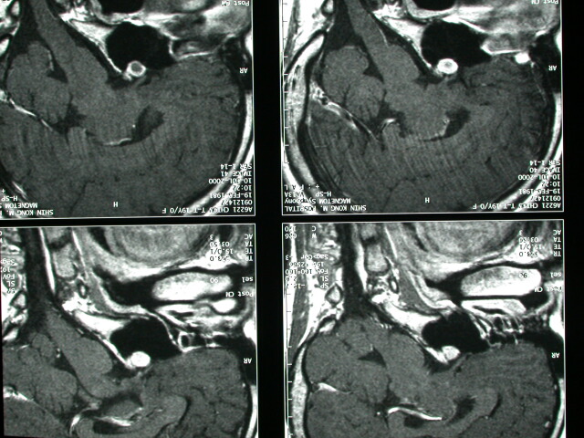
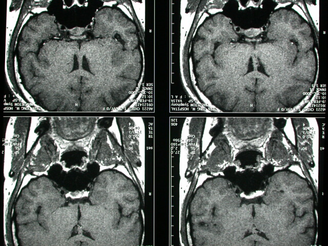
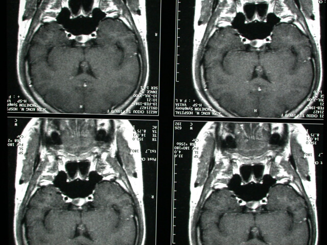
-Bulging of L't pit. gland with a 6mm delayed enhancing tumor in L't
pit. gland.
-The opitc chiasm seems not
-Preservation of the normal high SI of the neurohypophysis post. lobe)
on T1WI.
Diagnosis :
Microadenoma, L't pit. gland
Discussion :
Pituitary Microadenoma
=very small adenomas <10 mm
-usually become clinically apparent by hormone production (20-30%
of all pituitary
-prolactin elevation (>25 ng/mL in females)
-4 x 8 normal:adenoma demonstrated in 71%
->8 x normal:adenoma demonstrated in
-incidentaloma = nonfunctioning microadenoma / pituitary cyst
-NO imaging features to distinguish between different types o adenomas
MRI:
-small mass of hypointensity on pre- and postcontrast T1WI (nonenhancing)
-occasionally isointense on precontrast images + hyperintense on
postcontrast images
-enhancement on delayed images
-focal bulge on surface of gland
-focal depression of sellar floor
-deviation of pituitary stalk |


