History :
This 33 years old gentleman suffered from seizure attack (tonic-clonic
type)
Image finding :
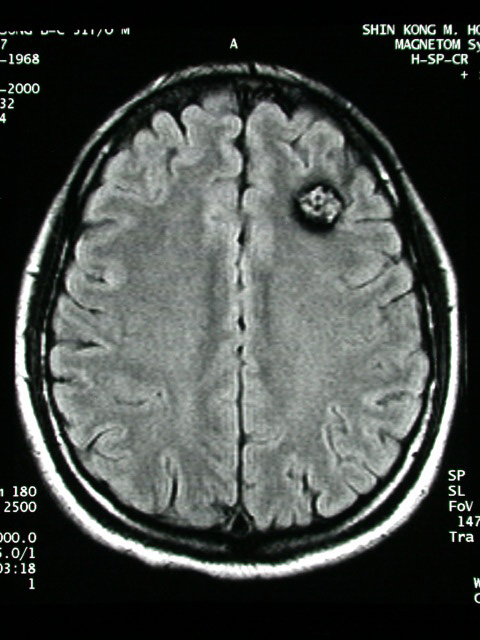
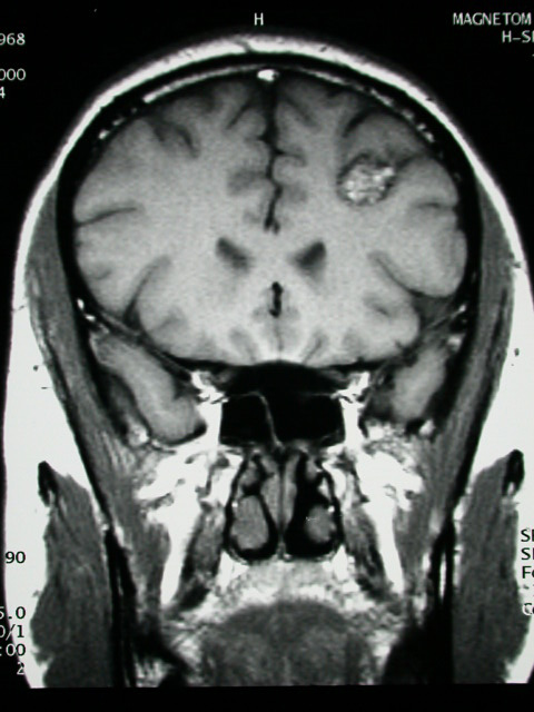
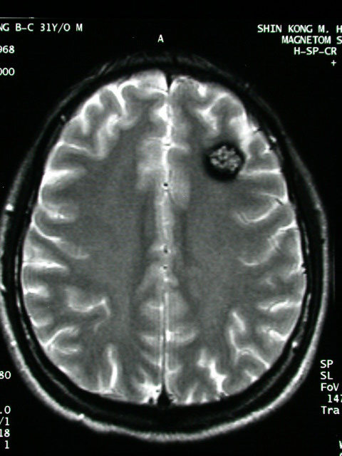
-Nodular lesion at L't frontal lobe.
-Dark signal intensity on both T1 & T2WI in the lesion may due
to hemosiderine deposition or calcification.
-Focal high signal on T1WI & low on T2WI of the lesion may be
calcified component.
Diagnosis :
Cavenous angioma, left frontal
Dicussion :
CAVERNOUS HEMANGIOMA OF BRAIN
= CAVERNOUS ANGIOMA = CAVERNOMA
Path:
-well-circumscribed nodule of honeycomblike large sinusoidal vascular
spaces separated by fibrous collagenous bands without intervening
neural tissue; slow blood flow in vascular channels
Age:
3rd-6th decade; M > F seizures (commonly presenting symptom)
Location:
cerebrum (mainly subcortical) > pons > cerebellum; solitary
> multiple NO obvious mass effect / edema
NCCT:
-extensive calcifications = hemangioma calcificans (20%)
-small round hyperdense region (CLUE)
-minimal surrounding edema
CECT:
-minimal / intense enhancement
-low-attenuation areas due to thrombosed portions
MR:
-well-defined area of mixed signal intensity centrally (= "mulberry"-shaped
lesion) with a mixture of
--increased signal intensity (= extracellular methemoglobin / slow
blood flow / thrombosis)
--decreased intensity (= deoxyhemoglobin / intracellular methemoglobin
/ hemosiderin / calcification)
-surrounded by hypointense rim (= hemosiderin) on T2WI
Angio:
-negative = "cryptic / occult vascular malformation"
Cx:
hemorrhage of varying ages
DDx:
-(1)Hemorrhagic neoplasm (edema, mass
-(2)Small AVM (thrombosed / small feeding vessels, associated hemorrhage)
-(3)Capillary angioma (no difference)
|


