History :
This 67 years old gentleman
Image finding :
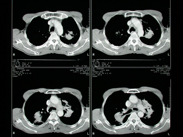
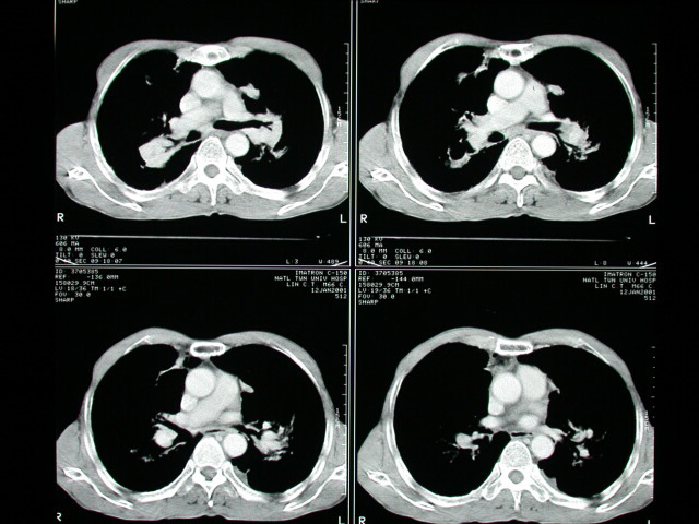
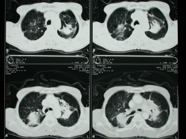
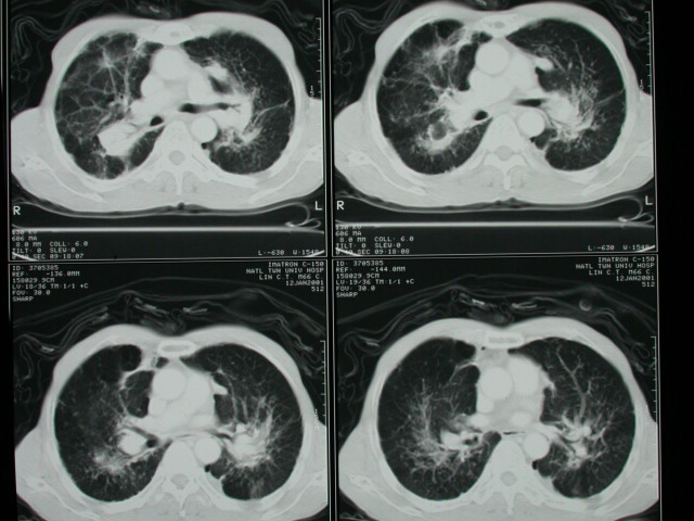
-A lipomatous lesion is noted in the right post. Back.
-Multiple linear and dot like lesions are noted at bil. Lungs. Pneumoconiosis
is compatible.
-Small amount of the pneumothorax is noted at the left upper lung
and ventral aspect of the left lower lung.
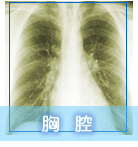
-Confluent radiopaque mass noted at bil. upper and middle lung fields.
-Progressive massive fibrosis is compatible.
-No definite evidence of pleural effusion.
.Diagnosis :
PNEUMOCONIOSIS
Discussion :
COAL WORKERS PNEUMOCONIOSIS
=CWP = ANTHRACOSIS = ANTHRACOSILICOSIS
=coal dust inhalation taken up by alveolar macrophages, in part
cleared by mucociliary action (particle size >5 μ), in part deposited
around bronchioles + alveoli, coal dust in itself is inert, but
admixed silica is fibrogenic
Simple CWP
=aggregates of coal dust = coal macules (usually <3 mm)
-NO progression in absence of further exposure
Histo:
-development of reticulin fibers associated with bronchiolar dilatation
(focal emphysema) + bronchiolar artery stenosis (decreased capillary
perfusion)
*poor correlation between symptoms, physiologic findings + roentgenogram
*small round 1-5 mm opacities, frequently in upper lobes (radiographically
only seen through superposition after an exposure of >10 years)
*nodularity correlates with amount of collagen (NOT amount of coal
dust)
Cx :
-(1)Chronic obstructive bronchitis
-(2)Focal emphysema (3)Cor pulmonale
Pneumoconiosis With Mass
Anthracosilicosis with:
-1.Granuloma (histoplasmosis, TB, sarcoidosis)
-2.Bronchogenic carcinoma (incidence same as in general population)
-3.Metastasis
-4.Progressive massive fibrosis
-5.Caplan syndrome (rheumatoid nodules) |

