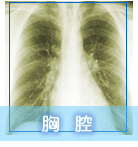
History :
This 83-year-old gentleman was suffered from dysphagia for 2wks and
fever for 3 days
Diagnosis :
Advanced esophageal cancer (poor differentiated carcinoma)
Discussion :
ESOPHAGEAL CANCER
-dysphagia (87-95%) of <6 months duration
-weight loss (71%)
-retrosternal pain (46%)
-regurgitation (29%)
Location:
-upper 1/3 (15-20%);
-middle 1/3 (37-44%);
-lower 1/3 (38-43%)
Radiologic types:
-(1)Polypoid / fungating form (most common)
--sessile / pedunculated tumor with lobulated surface
--protruding, irregular, polycyclic, overhanging, steplike "apple
core" lesion
-(2)Ulcerating form
--large ulcer niche within bulging mass
-(3)Infiltrating form
--gradual narrowing with smooth transition (DDx: benign stricture)
-(4)Varicoid form = superficial spreading carcinoma
Histo:longitudinal extension within wall without invasion beyond
mucosa / submucosa tiny confluent nodules / plaques
DDx:Candida esophagitis
Metastases:
(a)lymphogenic: anterior jugular chain + supraclavicular nodes (primary
in upper 1/3); paraesophageal + subdiaphragmatic nodes (primary
in middle 1/3); mediastinal + paracardial + celiac trunk nodes (primary
in lower 1/3)
(b)hematogenous: lung, liver, adrenal gland
CXR:
-widened azygoesophageal recess with convexity toward right lung
(in 30% of distal + midesophageal cancers)
-thickening of posterior tracheal stripe + right paratracheal stripe
>4 mm (if tumor located in upper third of esophagus)
-widened mediastinum
-tracheal deviation
-posterior tracheal indentation / mass
-retrocardiac mass
-esophageal air-fluid level
-lobulated mass extending into gastric air bubble
-repeated aspiration pneumonia (with tracheoesophageal fistula) |


