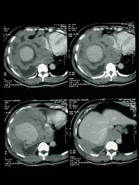
History :
The 60-year-old male was suffered from exertional dyspnea for 1 month.
Image finding :
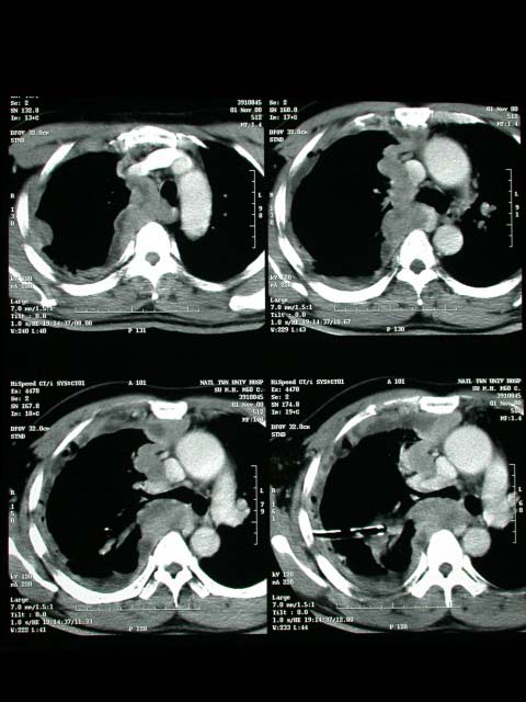
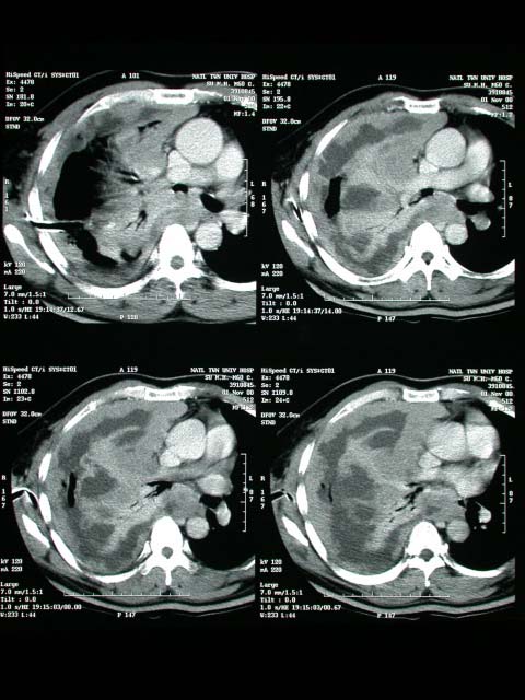
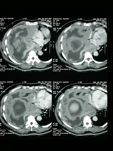
-Diffuse thickening and tumor infiltration of pleural space of right
lung.
-The tumor shows marked thickening of pleura and nodular appearance
as well as mass infiltration.
-There is compression of right lung associated with irregular lung
surface.
-Tumor involvement of the lung parenchyma esp. at right middle lobe
and right lower lobe is noted.
-There is encasement of RULB,right intermedial bronchus and obstruction
of RMLB and RLLB. Mild air bronchogram of collapse RLL is noted.
-The picture is consistent with mesothelioma of right pleural space
associated with lung invasion.
-There is also tumor involvement on the lymphadenopathy at right mediastinum.
-The SVC is displaced anteriorly and tumor infiltration at subcarinal
region and invasion to the right pericardium are also found. The mediastinum
is displaced toward left side.
Diagnosis :
Mesothelioma
Discussion :
Malignant Mesothelioma =DIFFUSE MALIGNANT MESOTHELIOMA =most common
primary neoplasm of pleura
Spread:
-(a)contiguous: chest wall, mediastinum, contralateral chest, pericardium,
diaphragm, peritoneal cavity; lymphatics, blood -
(b)lymphatic: hilar + mediastinal (40%), celiac (8%), axillary +
supraclavicular (1%), cervical nodes
-(c)hematogenous: lung, liver, kidney, adrenal gland
-extensive irregular lobulated bulky pleural-based masses typically
>5 cm / pleural thickening (60%)
-exudative / hemorrhagic unilateral pleural effusion (30-60-80%)
without mediastinal shift ("frozen hemithorax" = fixation
by pleural rind of neoplastic tissue); effusion contains hyaluronic
acid in 80-100%; bilateral effusions (in 10%)
-distinct pleural mass without effusion (<25%)
-associated with pleural plaques in 50% = pathologic HALLMARK of
asbestos exposure
-pleural calcifications (20%)
-circumferential encasement = involvement of all pleural surfaces
(mediastinum, pericardium, fissures) as late manifestation
-extension into interlobar fissures (40-86%) rib destruction in
20% (in advanced disease)
-ascites (peritoneum involved in 35%)
CT:
-pleural thickening (92%)
-thickening of interlobar fissure (86%)
-pleural effusion (74%)
-contraction of affected hemithorax (42%):
--ipsilateral mediastinal shift
--narrowed intercostal spaces
--elevation of ipsilateral hemidiaphragm
-calcified pleural plaques (20%)
MR (best modality to determine resectability):
-minimally hyperintense relative to muscle on T1WI
-moderately hyperintense relative to muscle on T2WI
DDx:
pleural fibrosis from infection (TB, fungal, actinomycosis), fibrothorax,
empyema, metastatic adenocarcinoma (differentiation impossible) |

