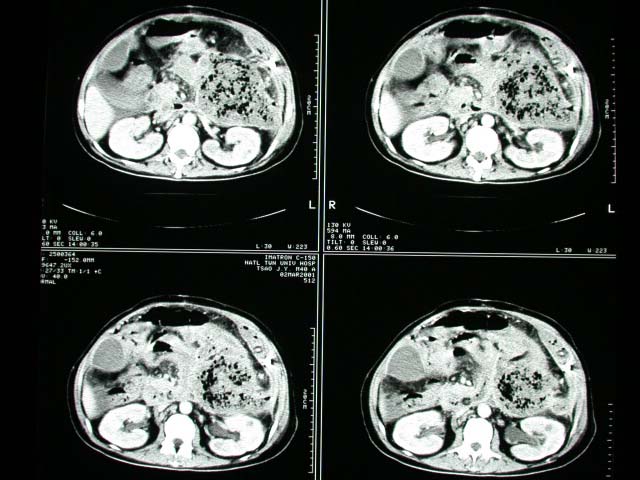
History :
This 40 y/o gentalman had abdominal distension and pancreatic enzyme
elevation. Dehydration and electrolyte imbalance was also noted.
Image finding :
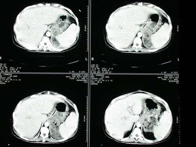
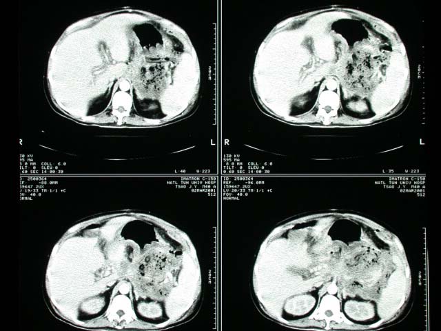
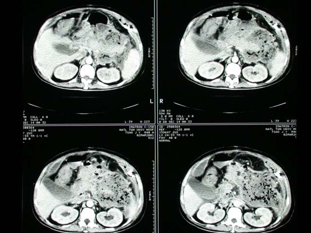
Abdominal CT showed pancreatic pseudocyst formation and infected necrotizing
pancreatitis was impressed.
Diagnosis :
Acute necrotizing pancreatitis with abscess formation
Discussion :
NECROTIZING PANCREATITIS: proteolytic destruction of pancreatic
parenchyma; mortality rate of 80-90%
HEMORRHAGIC PANCREATITIS: + fat necrosis and hemorrhage (b)SUPPURATIVE
PANCREATITIS: + bacterial infection
A. Diffuse pancreatitis (52%)
B. Focal pancreatitis (48%): location of head:tail = 3:2
Abdominal film:
"colon cutoff" sign = dilated transverse colon with abrupt
change to a gasless descending colon (inflammation via phrenicocolic
ligament causes spasm + obstruction at the splenic flexure impinging
on a paralytic colon)
CT findings in pancretitis (pancreatic visualization in 98%):
-no detectable change in size / appearance (29%)
-hypodense (5-20 HU) mass in phlegmonous pancreatitis; may persist
long after complete recovery
-hyperdense areas (50-70 HU) in hemorrhagic pancreatitis for 24-48
hours
-enlargement with convex margins + indistinctness of gland with
parenchymal inhomogeneity
-thickening of anterior pararenal fascia
-non-contrast-enhancing parenchyma during bolus injection (= pancreatic
necrosis) |

