 |
Chordoma, Sacrococcyx |
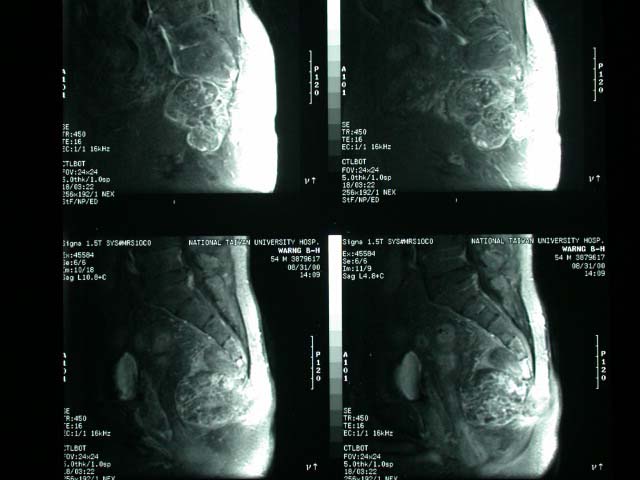
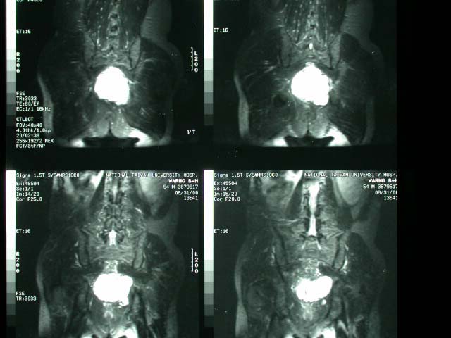
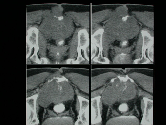
| History : A 54 y/o gentalman found to have a mass on the right buttock for 7 months. It was painful and may radiate to right thigh and lower leg. Image finding : MRI revealed a obulated mass arising from the sacrum coccyx junction. Pelvic CT with contrast enhancement shows : A large lobulated mass lesion located at the presacral space and extending to surrounding coccyx with destruction of the sacral bone. Calcified matrix is identified within this lesion. Diagnosis : Chordoma, Sacrococcyx Discussion : Sacrococcygeal Chordoma (50-70% of all chordoma) 40% of all sacral tumors Peak age: 40-60 years; M:F = 2:1 *low back pain (70%) *constipation / fecal incontinence *rectal bleeding (42%) sciatica *frequency, urgency, straining on micturition *sacral mass (17%) Location:esp. in 4th + 5th sacral segment -presacral mass with average size of 10 cm extending superiorly + inferiorly; rarely posterior location -displacement of rectum + bladder -solid tumor with cystic areas (in 50%) -osteolytic midline mass in sacrum + coccyx -amorphous peripheral calcifications (15-89%) -secondary bone sclerosis in tumor periphery (50%) -honeycomb pattern with trabeculations (10-15%) -may cross sacroiliac joint Prognosis: 8-10 years average survival; 66% 5-year survival rate (adulthood) DDx: Giant cell tumor, plasmacytoma, lymphoma, metastatic adenocarcinoma, aneurysmal bone cyst, atypical hemangioma, chondrosarcoma, osteomyelitis, ependymoma CT: -low-attenuation within soft-tissue mass (due to myxoid-type tissue) -higher attenuation fibrous pseudocapsule MR (modality of choice): -heterogeneous low to intermediate intensity on T1WI, occasionally hyperintense (due to high protein content) -very high signal intensity on T2WI (similar to nucleus pulposus with high water content) NUC: -cold lesion on bone scan -no uptake on gallium scan Prognosis: almost 100% recurrence rate despite radical surgery |
|


