 |
TCC |
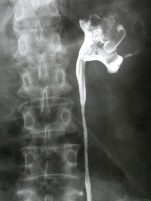
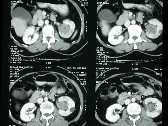
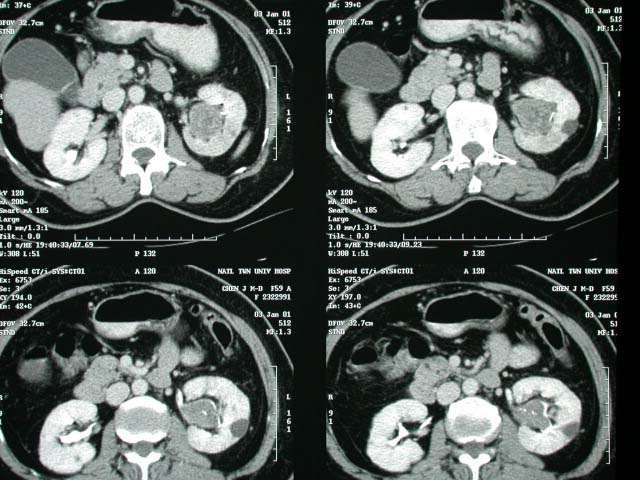
| History : This 59 y/o lady began to aware of frequency and dysuria since 2 months ago. Image finding : -The left upper calyces is not opacified suspect filling defect at left pelvis R/O tumor or blood clot. -Abnormal heterogeneous soft tissue mass filling inside the collecting system of left kidney including upper renal calyces and renal pelvis. -There is abnormal enhancement of kidney parenchyma at upper part of left kidney. Diagnosis : TCC Discussion : Renal TCC Site: extrarenal part of renal pelvis > infundibulocaliceal region IVP: -single / multiple filling defects in renal pelvis (35%) -"stipple sign" = contrast material trapped in interstices (DDx: blood clot, fungus ball) -dilated calyx with filling defect (26%) due to partial / complete obstruction of infundibulum --"phantom calyx" = failure to opacify from obstruction --±focal delayed increasingly dense nephrogram --"oncocalyx" = caliceal distension with tumor -caliceal amputation (19%) -absent / decreased excretion with renal atrophy (13%) due to long-standing obstruction of ureteropelvic junction -hydronephrosis with renal enlargement (6%) due to tumor obstruction of ureteropelvic junction US: -bulky hypoechoic (similar to renal parenchyma) mass lesion -splitting / separation of central renal sinus complex -infiltrative without bulge of renal contour -focal caliceal dilatation CT (52% accuracy due to overstaging): -sessile filling defect in opacified collecting system -thickening + induration of pelvicaliceal wall -central solid mass in renal pelvis expanding centrifugally -compression of renal sinus fat -invasion of renal parenchyma (infiltrating growth pattern) with preservation of renal contour -coarse punctate calcific deposits (0.7-6.7%) may mimic urinary calculi -variable enhancement of tumor |
|


